

In May of this year I wrote a post on modernizing variolation—the use of current science and modern tools to update the historical practice of variolation. In that post, I described my first use of hydrogen peroxide (H2O2) partial inactivation of a respiratory pathogen in nasal mucus from an infected donor (my brother) living in my home. My daughter and wife also became sick. So, the three other people I lived with under the same roof fell ill at about the same time. I did not get sick.
I had already sketched out all key steps of the “modernized variolation” (MV) procedures, and had purchased all necessary equipment, so I was ready to act. I have occasionally avoided being infected by sick people living in my home but only once since my daughter was born 4 ½ years ago. I was delighted to have avoided their illness; it seemed like a nasty bug and my brother stayed miserable for over a week. But I had taken other precautions to not get sick, so I waited for the next illness to try the procedure again before being tempted to declare a small victory.
Before I proceed with an account of the next, slightly larger victory, let’s back up a moment to review some basic information on variolation and the general approaches we are using at Radvac to modernize the traditional practices.
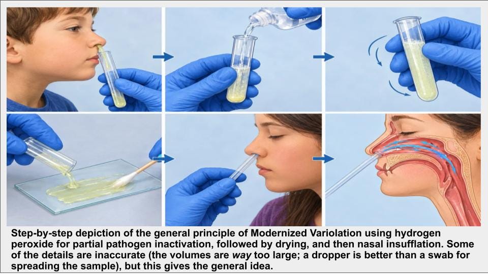
Variolation
Variolation was developed in response to smallpox by deliberately infecting people with a low dose of an attenuated form of the pathogen. The general procedure involved exposing a person to material taken from an infected patient (or recently variolated person), with the hope that the resulting infection would be mild yet provide robust immunity to the virus. Often, this was done through a process called inoculation—by rubbing smallpox scabs or fluid from pustules into scratches or incisions in the recipient’s skin.
While the smallpox virus was normally spread through the air and caused a severe infection starting in the lungs, infection by inoculation through the skin usually led to a milder infection that still induced a protective immune response. The reason for this milder response is because pathogens have evolved to attack through specific routes of infection. They often infect certain cells through specific molecular receptors on the cell’s surface. Most such receptors are absent from many other cell types. As a result, viruses have limited repertoires of attack via atypical routes. When they are placed outside of their normal “tropism” (specific cell type and tissue environments), they are often attenuated and more easily battled by the immune system. Host cell receptors for smallpox and some other viruses are common to many host cell types, but even viruses that have broad tropism infect certain cell types more easily than others. This is why dermal variolation worked reasonably well against smallpox. It is a form of attenuation.
Another variolation approach was to age smallpox scabs, grind them up, and insufflate (snort) the powder. The aging of scabs (for days or even weeks) prior to inoculation helped reduce infectiousness and increased recipient survival. This worked because the active viral load in the inoculum was reduced, while the total amount of viral antigen remained the same. If the aging period is so long (weeks or longer) that all viruses in the sample cannot replicate, the result is complete inactivation. Drying and aging the sample for days likely results in partial inactivation.
Defining features of MV
There are some notable advantages and defining features of “modernized variolation” (MV) that differentiate it from traditional and dangerous variolation practices of the past. Some of these come directly from modern vaccine research, including scientifically validated approaches to complete inactivation (prevents replication), partial inactivation (reduces viral load), attenuation (impairs replication), and a wide selection of adjuvants (compounds that are added to vaccines to amplify the immune response) to ensure safe but sufficient immune stimulation. Hydrogen peroxide is one example of a modern tool that is used to create experimental vaccines by partial or complete inactivation.1
Another advantage of MV over traditional variolation is far deeper knowledge about a wide range of pathogens, and the specific tropism of each, and the use of that knowledge as a procedural guide. Inoculation into a tissue that is outside of a pathogen’s normal range or tropism2 is a form of attenuation, which I have named extratropic attenuation.
DEFINITION: Modernized Variolation (MV) is a form of variolation that involves some combination of pathogen inactivation (partial or complete), pathogen attenuation, and enhancement of immunogenicity by the use of one or more adjuvants. The optimal combination of these variables is informed by modern science and current evidence.
Ideally, both partial inactivation and extratropic attenuation are used together. This is because certain pathogens have broad tropism. Dermal variolation of smallpox without these additional measures harmed and killed a lot of people.
On their own, completely inactivated pathogens typically elicit weak immunity when used for mucosal immunization because only a small fraction of them typically get through initial layers of defenses, such as the highly effective mucociliary barrier that protects nasal and other mucosal tissues. If a pathogen isn’t able to replicate once it does break through these defenses, it typically is cleared rapidly before it can stimulate robust adaptive immunity. One certainly doesn’t want to take the chance of failing to elicit a protective response—especially when dealing with a highly virulent pathogen.
This is where adjuvants play an important role in MV. Most potent vaccines consist of either active but attenuated viruses, or one or more inactive immunogens plus an adjuvant. The challenge of modernizing variolation is to use current scientific knowledge about pathogens and their tropisms, the immune system, adjuvants, and vaccines to effectively update the historical practice of variolation. The goal is to safely stimulate a robust adaptive immune response (neutralizing antibodies and T-cells) but not become ill.
The advantages of MV versus vaccines
MV has several important advantages over other pathogen countermeasures, including vaccines. These include the following:
Convenience, Immediacy, and Safety: Allows for rapid inoculation without vaccination-related delays, and without having to visit a public location (physician, clinic, etc.) during an outbreak, risking additional exposure.
Target Pathogen Specificity: Potentially provides tailored protection against the exact pathogen variant circulating in your immediate environment.
Natural Immune Mimicry: Elicits an immune response highly similar to a natural infection, but without the accompanying illness.
Dual-Layer Immunity: Potential to stimulate mucosal immunity and systemic immunity.
Self-Administration: Utilizes simple, painless methods including intranasal, dental floss3, oral enteric capsule, or transdermal using a microneedle roller
In my first test of MV I only used the intranasal approach. After this first test, for each future test of MV, I decided to use one of the other approaches. For reasons I’ll explain later, I will use the intranasal approach initially, followed by one of the others. For my second test I planned to use intranasal followed by transdermal administration.
The second test of MV
My second opportunity arrived on September 1. My daughter was a bit cranky in the morning and my wife said that she didn’t seem her normal self at bedtime the night before. By the afternoon she was sneezing and her nose began to run. This post provides a second case study in the form a of first-person narrative.
As this narrative shows, for a second time I used the MV procedure and did not get sick. This was the second consecutive illness in our home that I avoided, which is unprecedented. Given additional experience and perspective on the use of MV, at the end of the narrative I provide a brief interpretation of the results and why I think MV provides a substantial barrier to respiratory illness. And I give a rationale for why nasal administration should be the first MV procedure, rather than one of the three other methods studied by Radvac. These other approaches can be used subsequently.
Case 2: September 1-10 , 2026
Case subject: Preston Estep, Radvac founder and Chief Scientist
Case topic: This case study is a first-person narrative describing the development and second use of MV. The two main highlights of this narrative are 1) this is the first time I used the transdermal protocol (MV PROCEDURE TD1.0; see the bottom of this post for details), and 2) this the second consecutive time I have used MV in response to an illness in my home, and have avoided becoming sick.
Summary: On Day 1 of my 4.5 year-old daughter showing signs of infection with a respiratory pathogen, I collected a nasal mucus sample from her. I used the hydrogen peroxide inactivation protocol for inactivation of the pathogen (add an equal volume of H2O2), and then this mixture was administered intranasally. Unlike the previous test in May of this year, this time I followed administration with adjuvant triple antibiotic ointment. I also used intranasal antibiotic ointment or cream every subsequent day. For the first time I also tested the transdermal administration approach using a microneedle roller to administer a second sample that was treated with an equal volume of hydrogen peroxide for 30 minutes prior to administration.
Outcomes and bottom line: This is the second time I have used Modernized Variolation protocols based on partial pathogen inactivation by hydrogen peroxide. Both times I have avoided infection with a respiratory pathogen that infected the others in my home.
Case timeline
Day 1 (Sept 1):4 In the morning I noticed that my 4.5 year old daughter was a bit cranky and her energy level was lower than normal. My wife said that she didn’t seem her normal self at bedtime the night before. In the early afternoon, she began to show overt signs of a respiratory illness. Her nose was running, she was sneezing, and coughing.
Sample collection: Soon after overt symptom onset I had my daughter blow her nose directly into a 5 mL graduated tube. I put gloves on both hands and held the tube under one nostril at a time, closing the other nostril with a gloved finger. The mucus was very sticky and stringy and would not stay in the tube, so I used a gloved finger to gather the mucus into the tube opening. I used arm centrifuging to get the mucus into the bottom of the tube. Some of it remained adhered to the sides of the tube but I estimated the volume of mucus to be about 0.5 mL. In this protocol rough estimates are fine. Using a graduated dropper I added 0.5 mL of fresh hydrogen peroxide, mixed it well with the dropper, and spread it out onto a small, clean ceramic plate (a clear plate works well, allowing you to see the thickness of the spread mixture). I put the plate on a table in a secure location with good air circulation, and left the sample to dry at room temperature (about 75 deg F) for about 2 hours. At room temperature hydrogen peroxide decomposes into water and oxygen, and the water evaporates. This process is greatly accelerated by catalases, peroxidases, and transition metals in nasal mucus. The sample dried to a dull, thin film on the plate. I used a single-edged razor blade to scrape it off the plate and to chop it into a granular powder.
Intranasal administration: I first blew my nose with a tissue and then administered saline spray and then blew my nose again. I repeated this procedure. My right nostril remained mostly blocked, so I administered the majority of the sample into my left nostril by insufflating the powdered sample through a straw. I followed a couple hours later by using a clean cotton swab to apply a pea-sized dollop of triple antibiotic ointment into each nostril. Unlike the first time I used this MV protocol, I did not experience severe congestion, only mild to moderate congestion in the left nostril. Also unlike the first time, I did not feel slightly ill or feverish later or the next day.
Additional precautions: I held my breath or wore a mask when in close proximity to other people in the house, opened windows, kept HEPA air purifiers running, and ran on low speed the kitchen cooktop fan that vents outside. I ran Germ Guardian ultraviolet air sanitizers in my daughter’s room and in my office and bedroom.
Day 2: I woke up feeling fine. Nasal congestion is moderate in the left nostril and still heavy in the right.
Sample collection and H2O2 inactivation: In the morning I collected more of my daughter’s nasal mucus (a bit less than 0.5 mL). Using a graduated dropper, I mixed it with an equal volume of hydrogen peroxide, incubated for about 30 minutes.
Transdermal administration: I administered the mixture with a dermal roller into the top of my left thigh according to the protocol TD1.0. I applied a layer of triple antibiotic ointment to the administration area and bandaged it. The dermal roller procedure went relatively smoothly. The administration area is a bit red but no discomfort. The administration area is about 2 x 3 inches, which is covered perfectly by a 3x4 inch bandage. I used a dermal roller with 540 titanium microneedles, each 1.0 mm long. The pressure I used for rolling was light to moderate.
Following the transdermal administration procedure I applied a nasal dose of triple antibiotic ointment. I did this to keep my nasal mucosal immune system stimulated. I checked the transdermal administration site later in the day and it appeared a bit blotchy and reddish but not excessively so. I continued all additional precautions.
In the evening I collected a sample and stored it for possible future identification of the pathogen.
Day 3: I feel fine. Nasal congestion is moderate in both nostrils. I decided to try a new tube of double antibiotic cream (Neosporin pain relief cream containing neomycin sulfate and polymyxin B sulfate). I applied a nasal dose of the cream. The transdermal administration site still appears reddish but about the same as yesterday. I changed the bandage and applied a new layer of triple antibiotic ointment. I continued all additional precautions.
Day 4: I feel fine. Nasal congestion is light in the left nostril and moderate moderate to heavy in the right nostril. I applied a nasal dose of double antibiotic cream. The transdermal administration site appears about the same as yesterday but maybe a bit less pink/red. I removed the bandage. I continued all additional precautions.
Day 5: I feel fine. Nasal congestion is light to moderate moderate in both nostrils. I applied a nasal dose of double antibiotic cream. The transdermal administration site appears about normal (pink/red is gone). I continued all additional precautions.
Days 6 and 7: I feel fine. Nasal congestion is variable. My daughter and wife have mild symptoms but claim to feel much better. I discontinued wearing a mask but retained other precautions.
Days 8 and 9: My nasal passages and throat feel a bit raw. Those symptoms resolved and I attribute them to gastric reflux, which I get occasionally.
Day 10: I feel fine and my wife and daughter are fully recovered.
Outcomes: Subsequent to the MV procedures I did not become ill, despite members of the household showing overt signs of respiratory infection for several days. I banked a sample in the freezer for possible future identification of the pathogen.
My interpretation of the results
This is the second time I have used Modernized Variolation protocols based on partial pathogen inactivation by hydrogen peroxide. Both times I have avoided infection with a respiratory pathogen that infected the others in my home. Although I cannot say for certain that the MV self-administrations protected me against the pathogen in our home, the procedures aren’t terribly difficult and the outcomes have been good. I was somewhat vigilant about social distancing, but I was in the same home as two overtly sick people. Plus, my daughter is a typical 4 year old and does not totally understand the social distancing concept!
One possibility is that MV administration triggered innate immunity and congestion in my nasal passages. Maybe that alone protected me during the peak infectious period. Over 90% of respiratory pathogens enter the body through the nasal mucosa and respiratory tract. Therefore, it makes sense to block this entry point first. After the positive outcome from my first test of this protocol I reasoned that the enhanced nasal congestion and delivery of the donor’s mixture of immune signaling factors (cytokines, chemokines, and interferons) create a formidable primary defense against a respiratory pathogen. Therefore, intranasal administration will always be the first MV approach I will use against a respiratory pathogen.
Even if innate immunity is playing a critically important role, MV has the potential to create long-lasting adaptive immunity.
This is the first time I used the transdermal approach, a form of extratropic attenuation. The procedure went smoothly using a dermal roller with 540 titanium microneedles, 1.0 mm long. The administration site showed signs of irritation and possible immune stimulation, but the site was pinkish-red and blotchy but not overtly swollen, and did not appear to have been infected by an adventitious pathogen. These second and third administrations might have provided additional adaptive immunization.
Future tests
For the third test of MV I will first use the intranasal protocol followed on subsequent days by the dental floss protocol. As of this writing, my daughter has already brought home yet another respiratory infection from preschool, so stay tuned for Part 3 of the MV series to find out how this turns out. Coming very soon.
MV details and protocols
Additional details and full protocols for MV can be found by clicking the link to the MV white paper on the Radvac website: https://radvac.org/white-papers
Acknowledgements
Thanks to Chris Buck @ Viruses Must Die for brainstorming about transdermal applications by dermal roller. And thanks to the Radvac team, especially Brian Delaney for detailed feedback and edits.
Amanna, I.J., Raué, H.P. and Slifka, M.K., 2012. Development of a new hydrogen peroxide–based vaccine platform. Nature medicine, 18(6), pp.974-979. https://pmc.ncbi.nlm.nih.gov/articles/PMC3506259/pdf/nihms418821.pdf
Similar to the administration of smallpox via a dermal rather than respiratory route, but informed by current scientific knowledge of the immune system and tropisms of common pathogens.
Ingrole, R.S., Shakya, A.K., Joshi, G., Lee, C.H., Nesovic, L.D., Compans, R.W. and Gill, H.S., 2026. Floss-based vaccination targets the gingival sulcus for mucosal and systemic immunization. Nature Biomedical Engineering, 10(2), pp.370-389.
Note that there is a mixture of past to present tense in this narrative. This is intentional to distinguish between notes that were made from memory from those that were made in real time.
Probably the best way to popularize this would be to host in-person events where people can get hands on practice.